
Breast cancer detection through MRI has reached a turning point, offering new prospects for enhancing the accuracy and efficiency of medical imaging. These advancements are largely due to developments in artificial intelligence (AI), particularly explainable AI models. A significant contribution from recent research led by Felipe Oviedo, PhD, at Microsoft’s AI for Good Lab, showcases an AI model that is setting new standards in breast tumor detection with unparalleled precision. This innovation is particularly promising in scenarios where traditional AI models struggle, such as datasets with low cancer prevalence. The enhanced capabilities of this AI model address challenges in breast cancer screening methods, offering greater reliability in environments where tumor detection is a rarity.
Enhancing MRI Capabilities with AI
Understanding the Necessity for AI
Magnetic Resonance Imaging (MRI) is widely recognized for its advanced sensitivity compared to conventional mammograms, but it comes with its own set of challenges, like increased costs and frequent false positives. Radiologists have been exploring the potential of integrating AI to boost these capabilities, aiming to increase diagnostic accuracy while reducing time and costs. Previous studies have shown some improvements with AI, but many models lack rigorous evaluation, particularly in populations with low cancer prevalence, and often fail in terms of interpretability. This gap highlights the necessity of an improved model that can provide insightful predictions amidst imbalanced datasets.
Explainable AI Model Dynamics
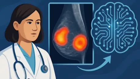
The explainable AI model applies an innovative mechanism to analyze breast MRI projections effectively. At its core is the deep explainable anomaly detection system known as the fully convolutional data description (FCDD) model. This system employs a convolutional neural network trained using an anomaly detection loss function. By producing a heat map of anomalous pixels, it can efficiently identify abnormalities and classify them as malignant or benign. The standout feature of this approach is its focus on detecting anomalies, contrasting with traditional binary classification techniques. This focus allows for robust learning of normal cases, ensuring that even minimal or variable malignant conditions are accurately identified.
Development and Evaluation of the New Model
Comprehensive Research Methodology
Spanning years of dedicated research, the development of this AI model involved a significant retrospective analysis of 9,738 breast MRI exams collected from a singular institution over 17 years, starting from 2005. These exams formed the foundation for the model’s development, specifically targeting malignancy detection in contrast-enhanced MRI scans. Evaluations of the model’s performance were conducted across three distinctive cohorts: a balanced detection group with 20% malignancy rates, an imbalanced group with only 1.85% malignancies, an internal independent test set comprising 171 exams, and a robust external dataset of 221 exams. This thorough approach ensured the model’s adaptability and performance across varying conditions.
Validation and Performance Metrics
The findings from these extensive evaluations revealed that the FCDD model consistently outperformed traditional binary cross-entropy models across different scenarios. Metrics like the area under the receiver operating characteristic curve (AUC) were pivotal in determining the effectiveness of the model. In balanced detection scenarios, the FCDD model achieved an AUC of 0.84, marking a significant improvement over the benchmark’s 0.81. For imbalanced scenarios, the model managed a competent AUC of 0.72 compared to the benchmark’s 0.69. Such results underscore the model’s adaptability and superior performance in diverse clinical settings, cementing its potential as a leading tool in breast cancer imaging.
Implications for Clinical Applications
Supporting Radiologists and Modifying Practices
These impressive results suggest that the innovative FCDD model could play a significant role in enhancing general breast MRI screening techniques. Notably, its application as a second reader for radiologists could revolutionize current practices, ensuring more accurate and quicker interpretations. According to Savannah Partridge, PhD, from the University of Washington, the model stands out due to its efficiency, leveraging minimal imaging information from detailed diagnostic exams. Its approach extends to both abbreviated MRI exams and full diagnostic ones, promising a reduction in both scanning times and interpretation requirements. Such efficiencies could greatly benefit clinical workflows.
Future of AI in Breast Cancer Diagnosis
Breast cancer detection using MRI technology has reached a pivotal stage, bringing fresh opportunities to improve the accuracy and efficiency of medical imaging. This progress is largely driven by breakthroughs in artificial intelligence (AI), specifically through explainable AI models. Recently, an impressive contribution to this field was made by Felipe Oviedo, PhD, from Microsoft’s AI for Good Lab. His team has developed an AI model that is redefining breast tumor detection, offering unmatched precision. This cutting-edge innovation stands out in situations where conventional AI models encounter challenges, such as in datasets with low cancer prevalence. By significantly enhancing the capabilities of breast cancer screening methods, this AI model provides increased reliability, especially in settings where detecting tumors is uncommon. These advancements promise to transform the landscape of breast cancer screening, making it more effective and reducing false negatives, thereby offering hope for earlier and more accurate diagnoses.